MRI is a non-invasive and non-radiating imaging technique, allowing to obtain high-resolution images ranging from array of soft tissues to different anatomical region of the body such as the brain. Different types of MRI sequences exist and can be used depending on the process or structure to be studied. Our research uses a multimodal MRI approach to characterize the brain tissues of patients with epilepsy, encompassing both structural and functional acquisitions.
A structural MRI sequence aiming at visualizing the locus coeruleus – a small brainstem nucleus involved in the antiseizure effects of VNS – has been developed and optimized in-house in our lab with the SIGNATM Premier 3T MRI system of Saint-Luc University Hospital. Part of our research focuses on evaluating the integrity of this brainstem nucleus and its potential impact on the therapeutic effectiveness of VNS.
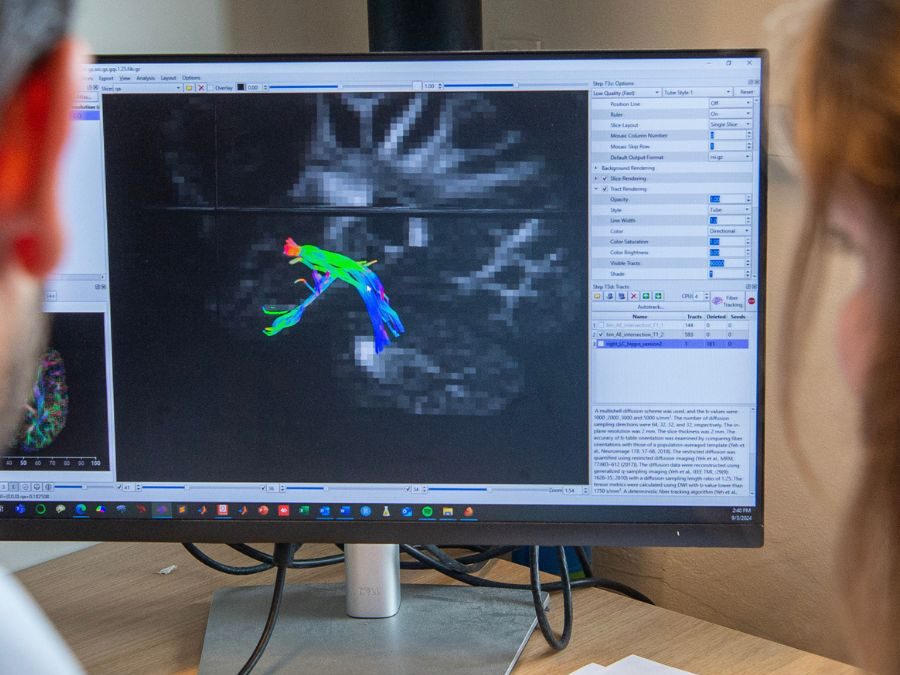
High-gradient multi-shell diffusion MRI was also used in our lab and constitutes a powerful imaging technique to evaluate white matter microstructure based on the diffusion of water molecules within the brain. In this context, our lab investigated how the integrity of various white matter tracts – including tracts projecting from the locus coeruleus to the hippocampus, and thalamocortical tracts – could be linked to the therapeutic effectiveness of VNS, using different (multi-compartment) diffusion models. These models included: Diffusion Tensor Imaging (DTI), Neurite Orientation Dispersion and Density Imaging (NODDI), distribution of anisotropic microstructural environments in diffusion-compartment imaging (DIAMOND), and Microstructure Fingerprinting (MF).
Functional MRI is used to measure and map brain activity in an array of clinical conditions. Using task-based paradigms, changes in cerebral blood flow occur in response to neural activity in specific brain regions involved in the task being performed.
Resting-state functional MRI is similar to the “classical” functional MRI in regard to its physical aspect (i.e. measuring the fluctuation of the blood flow in the different regions of the brain) but differs with the type of activity asked from the patient. The subject is asked to empty his mind and try to avoid thinking about anything, and the sequence records thus the activity of the brain at rest (hence the term resting-state). Thereafter, the signal is processed in order to identify the different regions that show a correlation between each other, depending on the purpose of the study.